Feline Infectious Peritonitis (FIP) is a complex and serious viral disease affecting cats. It is caused by a mutation of the feline coronavirus (FCoV). FIP remains a diagnostic challenge for veterinarians due to its complex pathogenesis, variable clinical presentation, and the difficulty in establishing a definitive diagnosis.
In this Vet Info, we review the recently published guidelines from the American Association of Feline Practitioners (AAFP) on the diagnosis of FIP.
General Considerations
An ante-mortem diagnosis of FIP should never be based on the results of a single diagnostic test.
In any cat suspected of having FIP, it is important to consider the signalment, clinical signs, and bloodwork findings, and to pursue additional diagnostic testing as appropriate (see below).
Antibody titers—whether measured in blood, cerebrospinal fluid, or aqueous humor—are not reliable for diagnosing FIP.
All cats suspected of FIP should undergo a complete blood workup. Additional testing depends on the clinical presentation and falls into the following categories:
- Cats with effusions
- Cats with neurological signs
- Cats with ocular signs
- Cats with nonspecific signs and no effusion
The diagnostic accuracy of tests is highest on effusion fluid. If effusion is present, it should always be examined. Repeated POCUS (point-of-care ultrasound) exams may be indicated to detect effusions.
General Blood Work (Hematology and Biochemistry)
Common hematologic abnormalities:
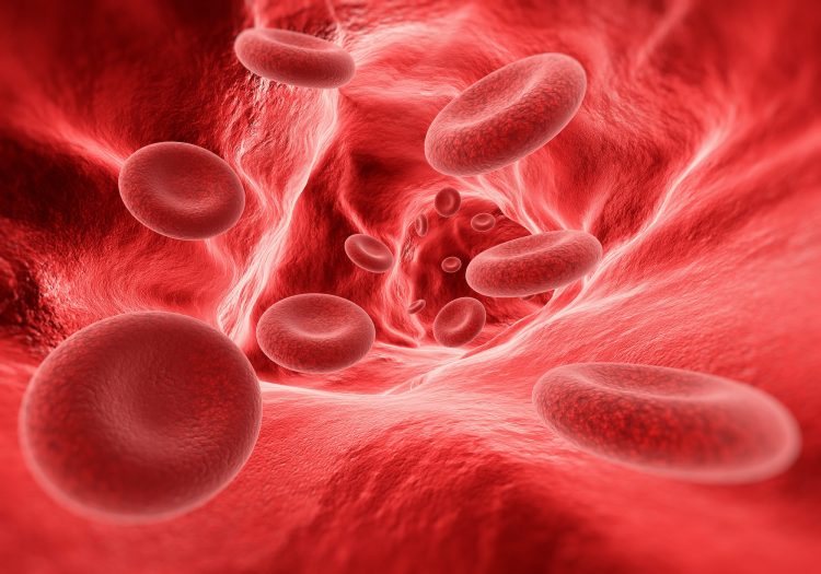
- Anemia (non-regenerative or regenerative if immune-mediated hemolysis is present), microcytosis (low MCV).
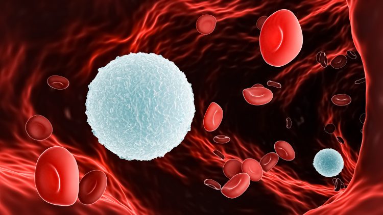
- Lymphopenia (especially in cats with effusions), neutrophilia (possibly with a left shift).
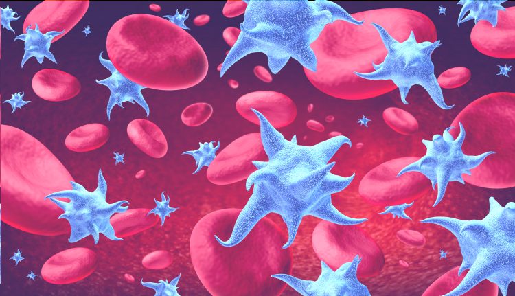
- Thrombocytopenia.
Common biochemical abnormalities:
- Hyperproteinemia and hyperglobulinemia (sometimes with normal total protein). Hyperglobulinemia—either monoclonal or polyclonal—occurs in 89% of FIP cases, but can also result from other inflammatory diseases. It is therefore not pathognomonic for FIP.
- An albumin:globulin (A:G) ratio < 0.4 may support a suspicion of FIP, but must always be interpreted in conjunction with other diagnostic findings. A ratio > 0.6–0.8 makes FIP less likely, but does not rule it out.
- Hyperbilirubinemia is frequently seen in cats with FIP and effusions.
- Azotemia and elevated liver enzymes may also be present.
A cat should never be diagnosed with FIP based solely on hematology or biochemistry results. These findings must always be interpreted alongside additional diagnostic criteria.
Additional Viral Detection Tests
Immunostaining (Immunocytochemistry, Immunofluorescence, Immunohistochemistry):
These techniques can be performed on:
- Effusion fluid (immunofluorescence staining).
- Fine-needle aspirates of tissues (e.g., mesenteric lymph nodes, liver, spleen) or cerebrospinal fluid (immunocytochemistry).
- Tissue biopsies (immunohistochemistry).
Immunohistochemistry on tissue biopsies (e.g., from mesenteric lymph nodes, intestinal wall, spleen, liver, kidney, omentum) is the gold standard for confirming FIP. Multiple tissue biopsies are recommended when samples are taken.
- Immunocytochemistry and immunofluorescence staining have low sensitivity (~50%) but high specificity (90–95%).
- A positive result makes FIP very likely.
- False positives can occur, particularly when testing CSF, aqueous humor, and—less commonly—effusion fluid.
Real-Time PCR (RT-PCR):
This quantitative test can be performed on blood, tissue aspirates or biopsies, effusion fluid, CSF, and aqueous humor. It detects the presence of enteric coronavirus RNA, which can be found in extraintestinal tissues of both FIP and non-FIP cats. Cats with FIP often have higher viral RNA loads, so quantitative PCR is recommended.
A mutation-specific RT-PCR test is also available, which looks for mutations (e.g., in the S gene coding for spike proteins) characteristic of the FIP virus. Performing the test on multiple tissues can improve sensitivity.
Some studies found this mutation-specific RT-PCR more specific than standard RT-PCR, but other studies reported false positives, and no clear superiority was demonstrated.
Additional Testing in Cats with Effusions
In male cats, scrotal effusion may be the only effusion present.
Typical features of FIP-associated effusion:
- High protein content (>35 g/L), often leading to clumping or string formation.
- Color ranges from straw-yellow to clear or turbid.
- Low cell count (<5 x 10⁹ cells/L), though it can occasionally be high.
Protein electrophoresis on the fluid has limited additional value.
Other causes of effusions should always be ruled out, using total protein, cell counts, and cytology. If sepsis is suspected, a culture of the fluid should be performed.
Rivalta’s test is a simple, inexpensive test that should always be considered:
How to perform:
- Fill a plastic tube (e.g., 10 ml) with 7 ml of distilled water (room temperature).
- Add 2–3 drops of vinegar and mix well.
- Place one drop of effusion fluid onto the surface.
- Positive: the drop remains suspended, keeps its shape while sinking, forms a jellyfish-like structure, or breaks into visible particles.
- Negative: the drop completely dissolves.
Results:
- Negative Rivalta’s: FIP is unlikely.
- Positive Rivalta’s: further testing (e.g., RT-PCR on effusion fluid) is recommended.
RT-PCR (if Rivalta’s is positive):
- Negative: FIP is unlikely, though false negatives can occur.
- Positive: FIP is likely, especially if the viral load is high.
Sensitivity increases if testing is also done on tissue samples (e.g., fine needle aspirates of mesenteric lymph nodes, spleen, liver).
Immunofluorescence staining:
- Low sensitivity, high specificity.
- A positive result supports a diagnosis of FIP.
- A negative result does not rule out the disease.
Additional Testing in Cats with Neurological Signs
These cats often lack effusions.
Cerebrospinal fluid (CSF) typically shows elevated protein levels and pleocytosis.
RT-PCR on CSF (and optionally on tissue aspirates of mesenteric lymph nodes, liver, spleen):
- Negative: FIP unlikely, but false negatives possible
- Positive: FIP likely, especially if viral load is high
Immunocytochemistry on CSF:
Only one study evaluated the reliability of this test, reporting a sensitivity of 85% and specificity of 83%.
Additional Testing in Cats with Ocular Signs (e.g., Uveitis)
Start with cytology of aqueous humor to exclude other causes, such as lymphoma.
RT-PCR on aqueous humor (and optionally on fine needle aspirates of mesenteric lymph nodes, liver, spleen):
- Negative: FIP unlikely, but false negatives possible.
- Positive: FIP likely (especially if viral load is high).
Additional Testing in Cats with Nonspecific Signs (No Effusion)
These are the most difficult to diagnose. Some of these cats may develop effusions later. Repeated POCUS exams may help.
Begin with RT-PCR on blood and tissue aspirates (mesenteric lymph nodes, spleen, liver)*
*Do not rely on blood alone—most FIP cats have low viral RNA in the blood, leading to false negatives.
- Negative: FIP unlikely. If clinical suspicion remains high, perform histopathology and immunohistochemistry on biopsies from mesenteric lymph nodes, spleen, and liver (via laparotomy or laparoscopy).
- Positive: FIP is very likely.
Immunocytochemistry on fine-needle aspirates (mesenteric lymph nodes, liver, spleen):
- Low sensitivity, high specificity.
- Positive result = FIP likely.
- Negative result = does not rule out FIP.
More Information
For more information: https://catvets.com/guidelines/practice-guidelines/fip-guidelines.
Voor een praktisch schematisch algoritme: https://catvets.com/public/PDFs/PracticeGuidelines/Guidelines/FIP/AAFP_EveryCat_Diagnostic_Wor
k_Up_of_FIP_Diagnostic_Approach.pdf
References
- Felten S and Hartmann K. Diagnosis of Feline Infectious Peritonitis: A Review of the Current Literature. Viruses 2019;11(11):1068
- Thayer V et al. 2022 AAFP/Every Cat Feline Infectious Peritonitis Diagnosis Guidelines. Journal of Feline Medicine and Surgery (2022)24:905-933