In collaboration with Dr. Adrien-Maxence Hespel from the University of Tennessee, we present the following radiology quiz. The goal of this case is to practice identifying radiographic findings and formulating a radiographic diagnosis based on a brief patient description. The final section provides additional information about the treatment.
In collaboration with Dr. Adrien-Maxence Hespel from the University of Tennessee, we present the following radiology quiz. The goal of this case is to practice identifying radiographic findings and formulating a radiographic diagnosis based on a brief patient description. The final section provides additional information about the treatment.
A 4-year-old spayed female Labrador Retriever is presented with fever of unknown origin (FUO).
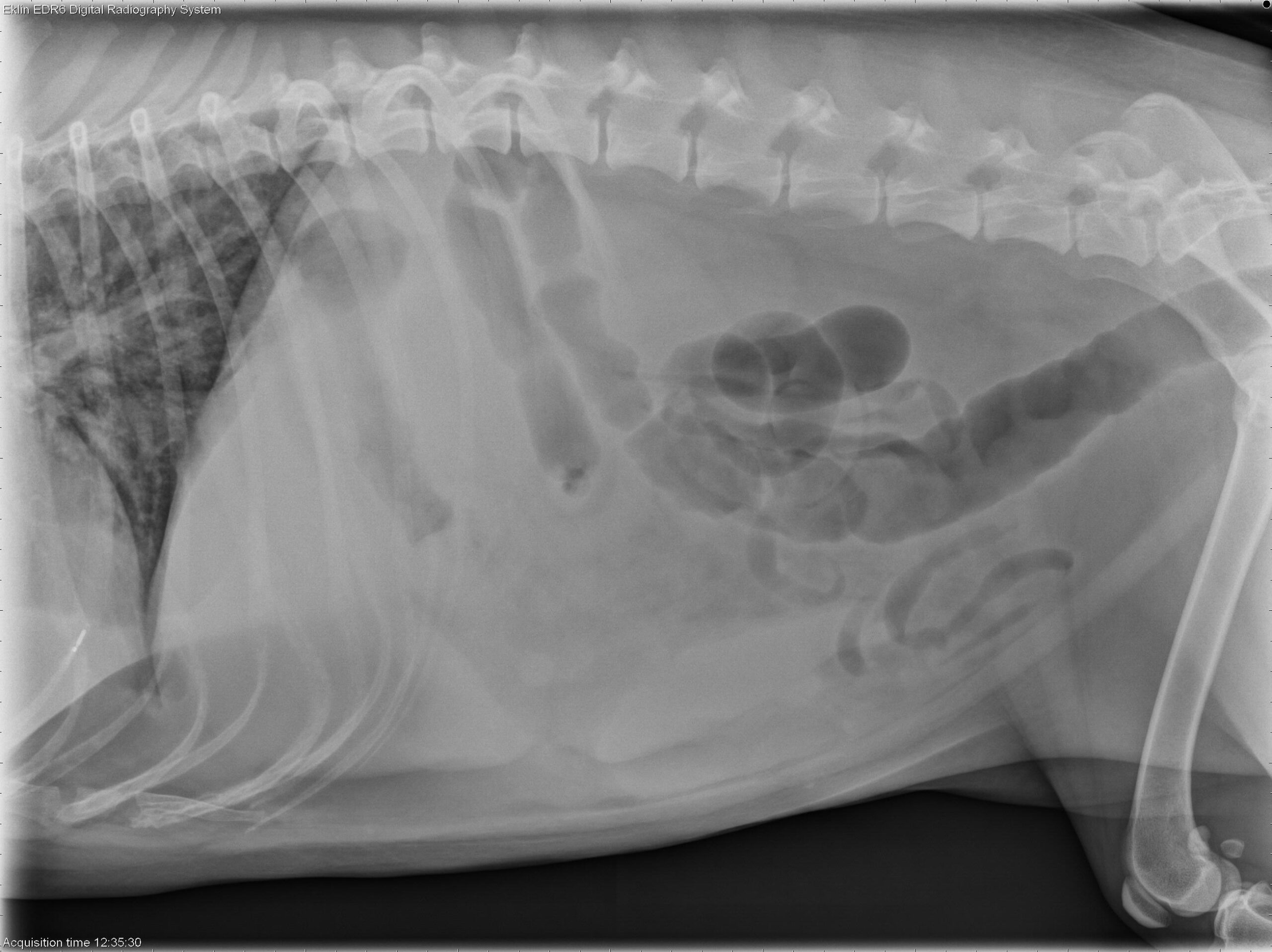
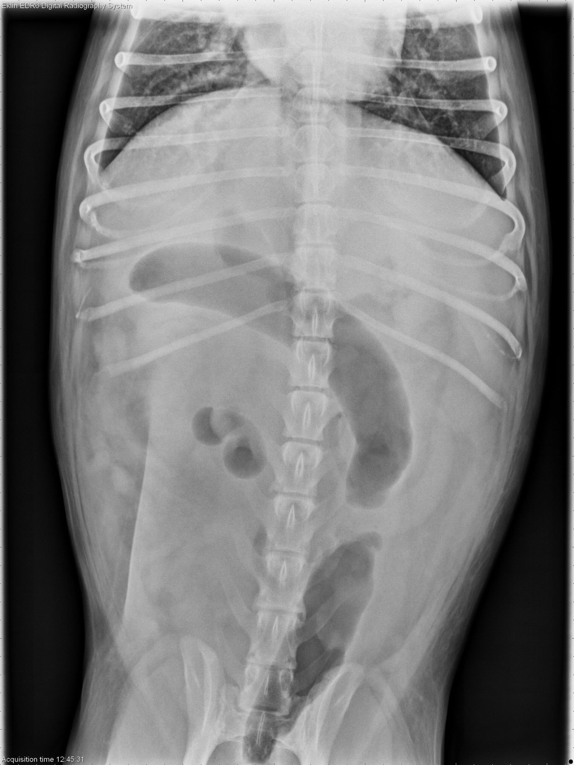
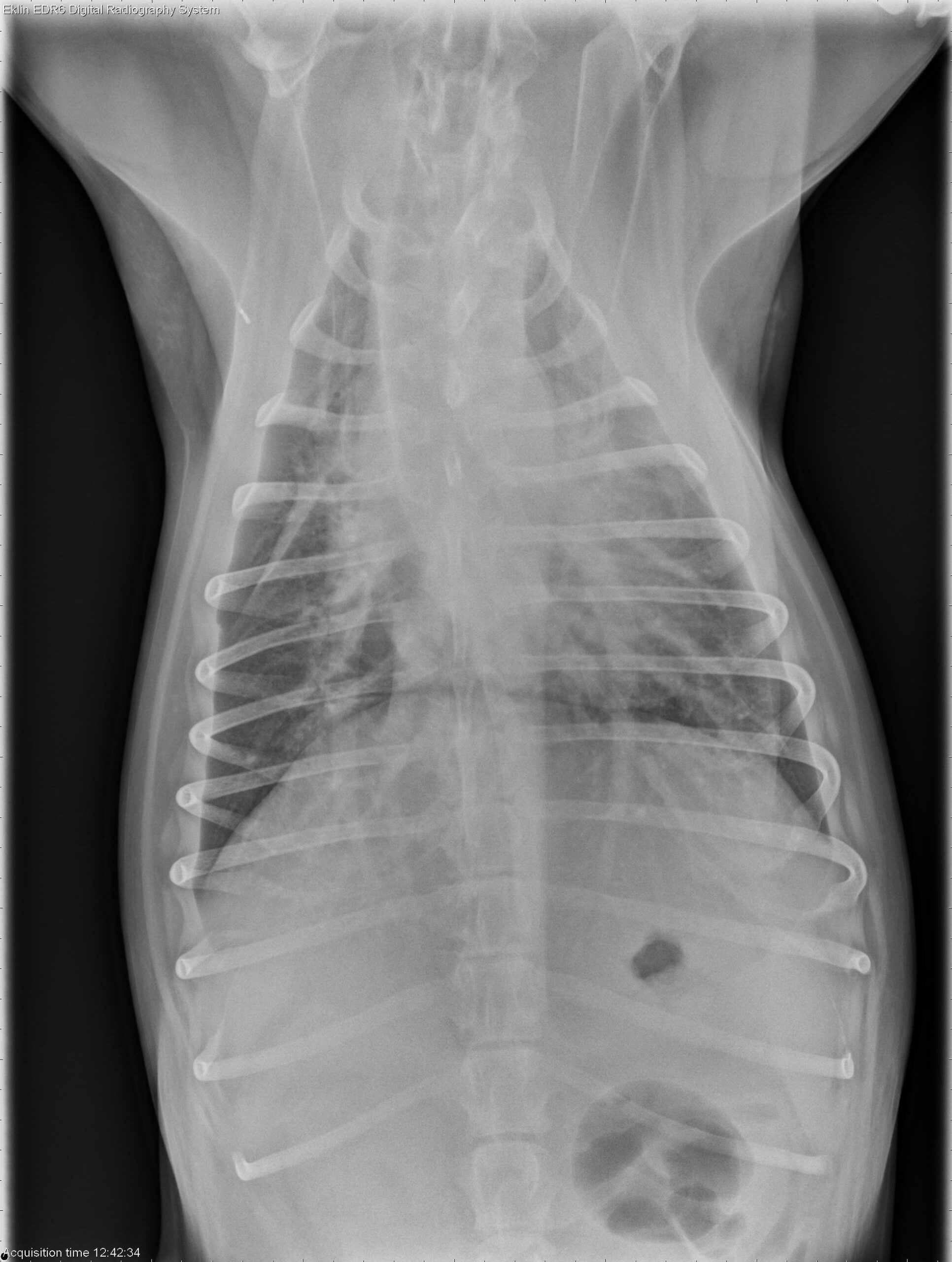
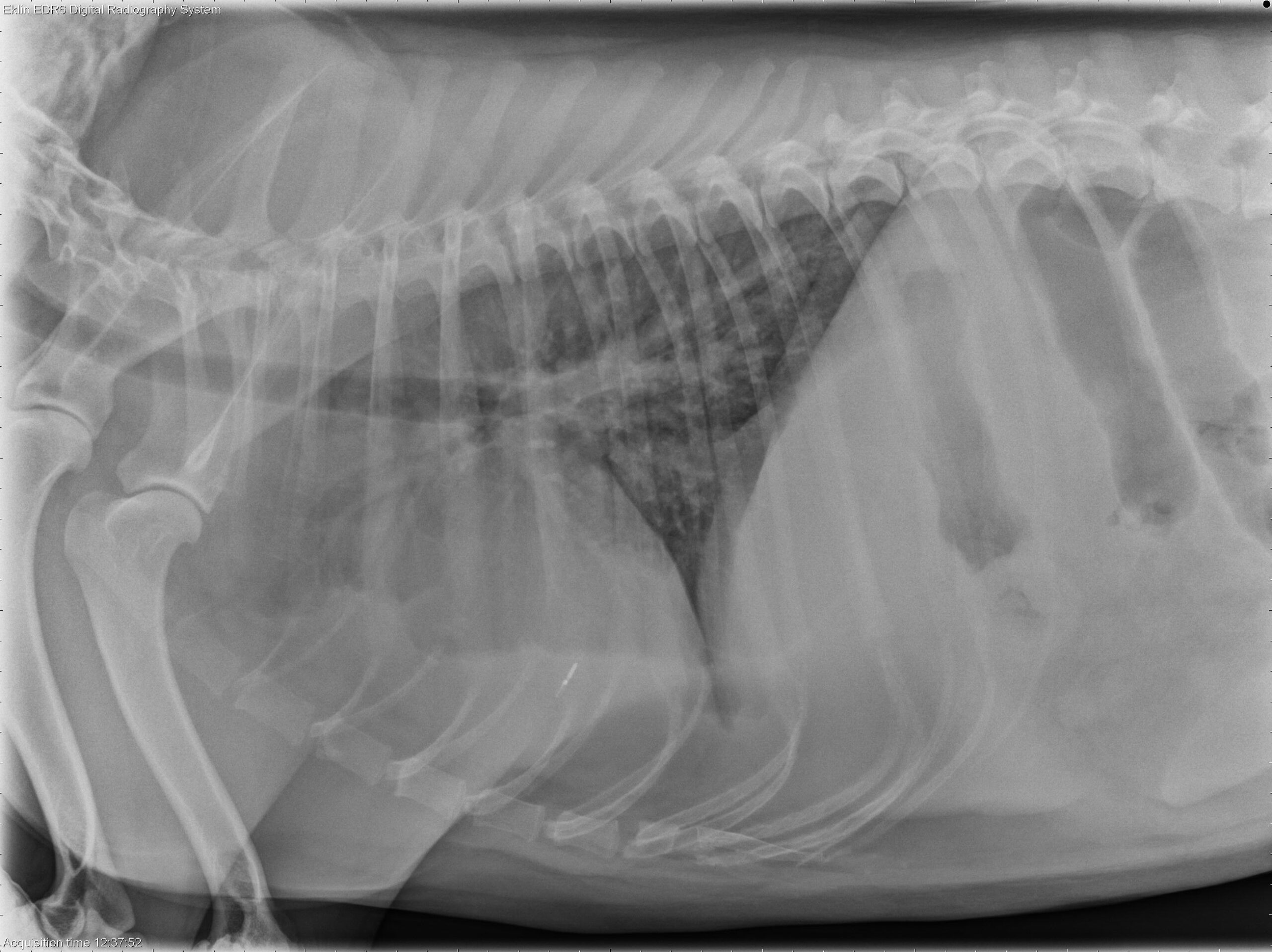
Below you’ll find the 4 X-ray images of the thorax. Use the arrows to scroll through, or click on the image to enlarge.
Thorax: There is increased soft tissue opacity within the pleural space, with retraction of the lung lobes and partial silhouetting with the cardiac silhouette, consistent with pleural effusion. A patchy broncho-interstitial pattern is present in the lungs. The cardiovascular structures appear normal.
Abdomen: The abdomen is distended with poor peritoneal detail. The liver and spleen are enlarged with rounded margins. The colon is moderately distended with gas. The small intestines appear normal. The urinary tract is poorly visualized.
Bicavitary effusion may result from round cell neoplasia, metabolic disorders, or inflammatory diseases. The hepatosplenomegaly is most suggestive of lymphoma. Differential diagnoses for the pulmonary pattern include lymphoma and non-cardiogenic pulmonary edema.
Ultrasound-guided fine-needle aspirates of the liver and spleen were obtained. Cytology confirmed hepatosplenic large granular cell lymphoma (NK-cell type). Thoracic POCUS revealed the presence of pleural effusion and B-lines within the lung parenchyma, indicative of pulmonary fluid accumulation. This suggests concurrent involvement of the lungs, most likely due to non-cardiogenic pulmonary edema.