In collaboration with Dr. Adrien-Maxence Hespel from the University of Tennessee, we present the following radiology quiz. The goal of this case is to practice identifying radiographic findings and formulating a radiographic diagnosis based on a brief patient description. The final section provides additional information about the treatment.
A 15-year-old neutered male Manx cat is presented with a history of two ‘episodes.’ The owner suspects these were epileptic seizures and describes them as “the cat became stiff and collapsed.” Cardiac auscultation reveals a grade 2/6 systolic murmur.
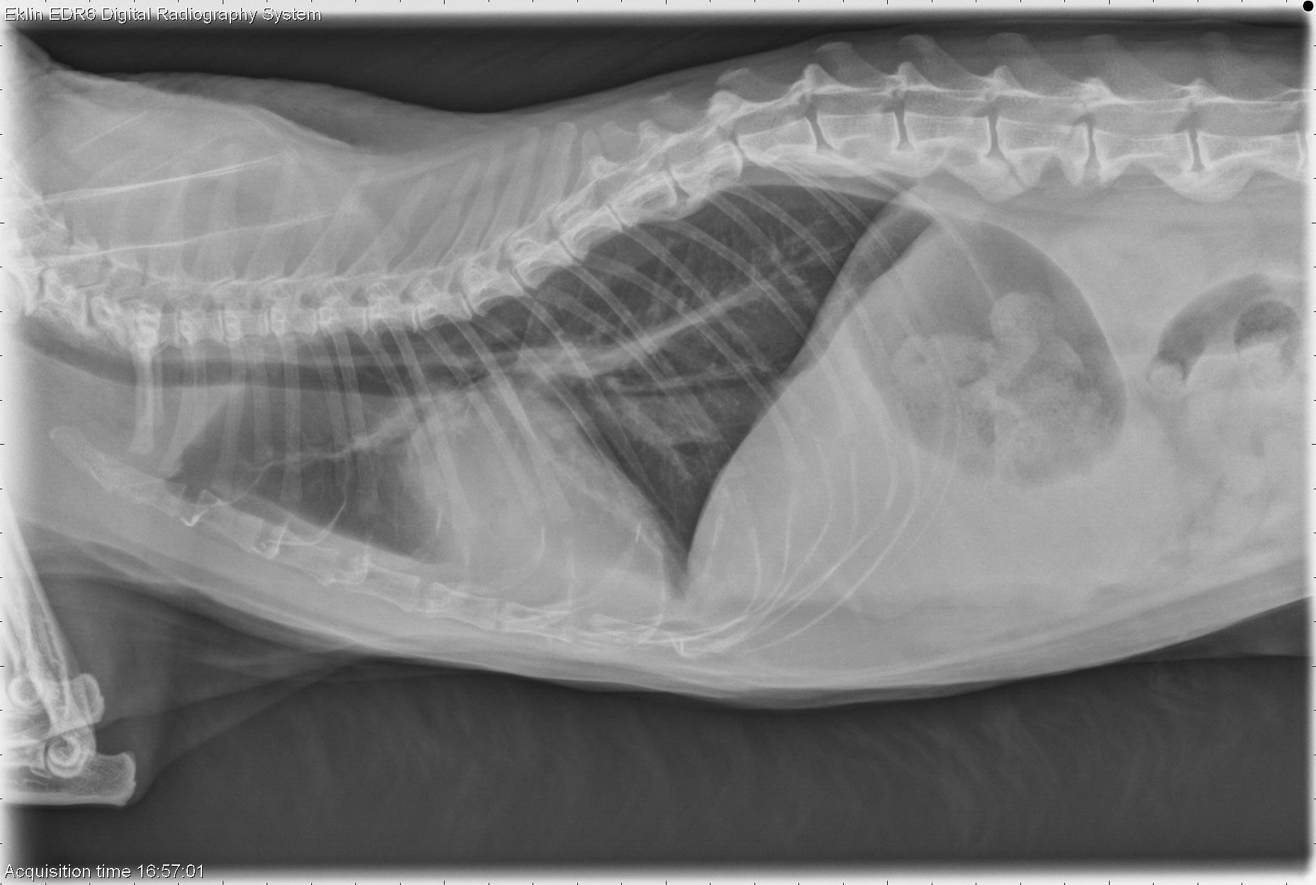
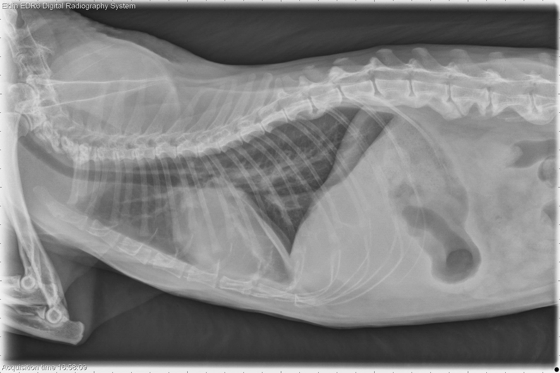
Below you’ll find the 3 X-ray images of the thorax. Use the arrows to scroll through, or click on the image to enlarge.
The cardiac silhouette is markedly enlarged on all projections. On the lateral view, the trachea is dorsally displaced, and on the dorsoventral projection, the cardiac silhouette occupies approximately three-quarters of the thoracic width. The pulmonary vessels are moderately enlarged. Several fissure lines are visible in the pleural space, indicative of pleural effusion. In the visible portion of the abdomen, there is poor serosal detail. Bridging spondylosis deformans is present in the lumbar spine.
Cardiomegaly (hypertrophic, restrictive, or dilated cardiomyopathy) with congestive heart failure.
Echocardiography revealed end-stage hypertrophic cardiomyopathy with congestive heart failure.