In collaboration with Dr. Adrien-Maxence Hespel from the University of Tennessee, we present the following radiology quiz. The goal of this case is to practice identifying radiographic findings and formulating a radiographic diagnosis based on a brief patient description. The final section provides additional information about the treatment.
10 year old female cat. Presented for anemia.
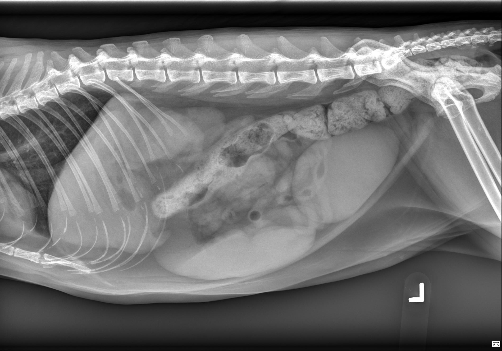
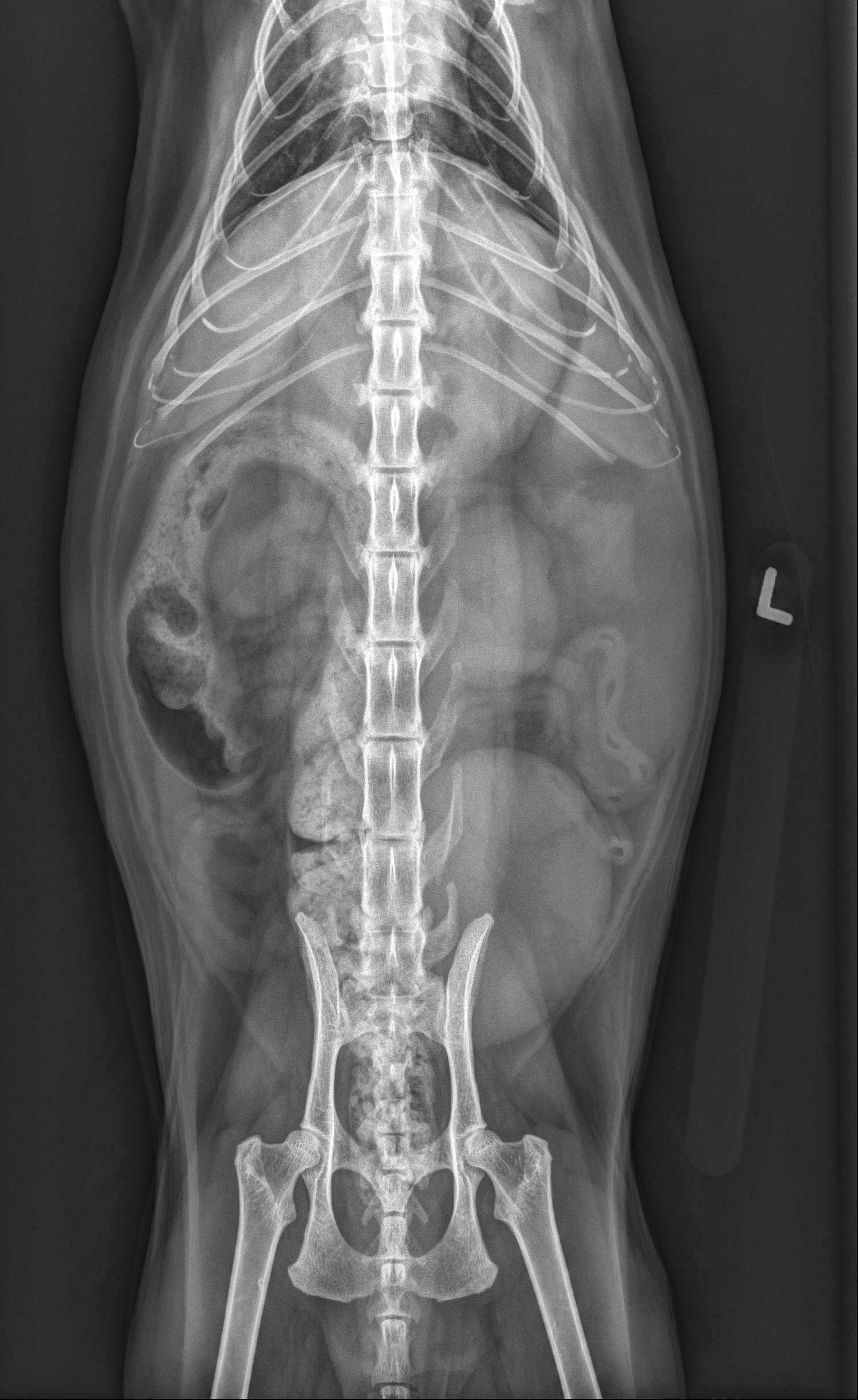
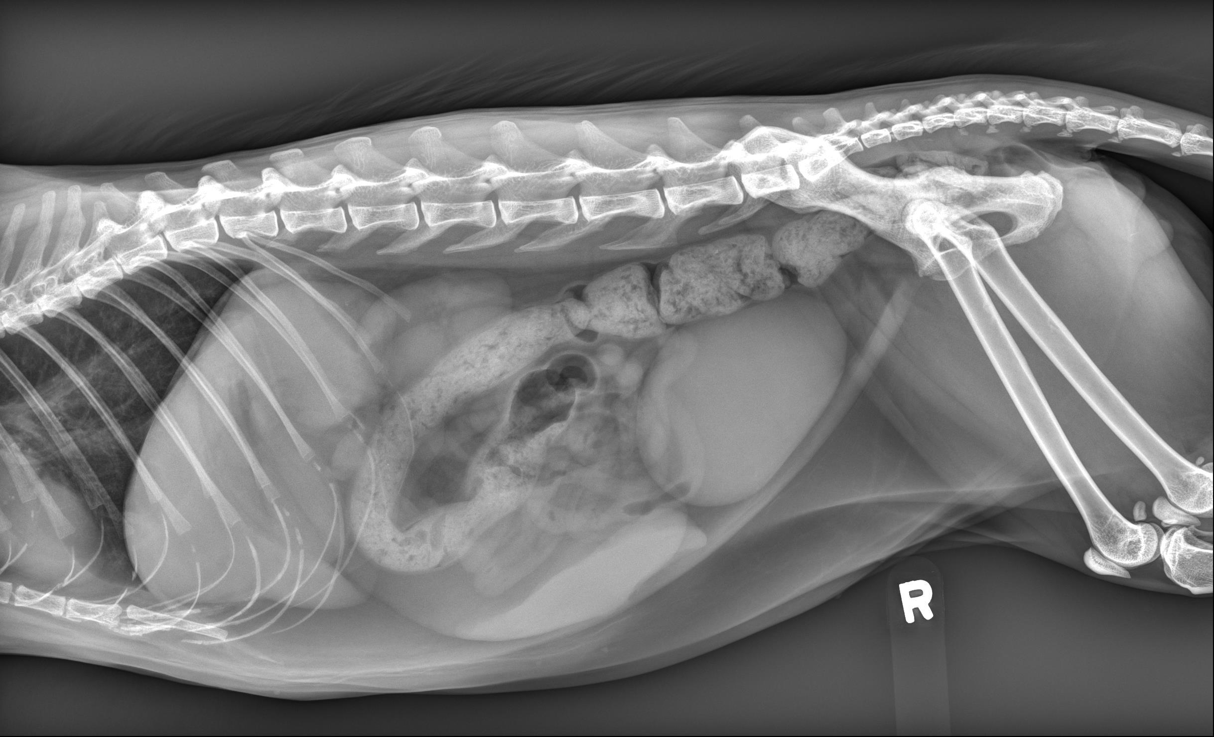
Below you’ll find the 3 X-ray images of the abdomen. Use the arrows to scroll through, or click on the image to enlarge.
Orthogonal radiographs of the abdomen are available for interpretation.
The abdominal detail is within normal limits.
The spleen is severely enlarged and slightly irregularly marginated on all 3 views. Equivocal hepatomegaly.
The left kidney is mildly irregularly marginated. The right kidney is smaller than the left kidney and also has blunt margination.
On the left lateral projection, ventral to the colon in the mid abdomen there is the impression of soft tissue mass. However this is not correlated on any other projection and is likely to represent summation artifact of normal abdominal content.
Severe splenomegaly. This is concerning for underlying infiltrative disease such as lymphoma.
The FNAs of the spleen were indicative of possible lymphoma.