Thorax, 3 views:
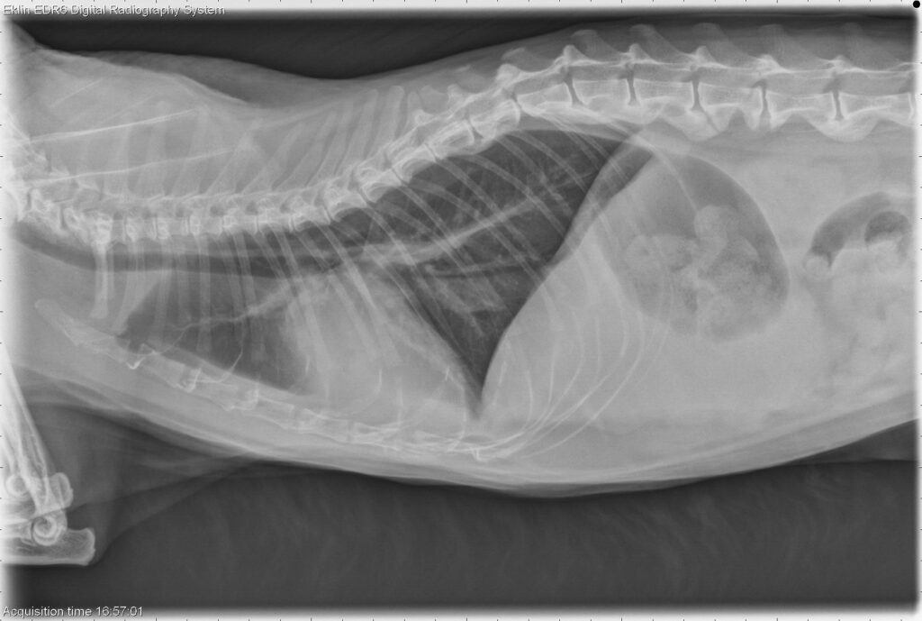
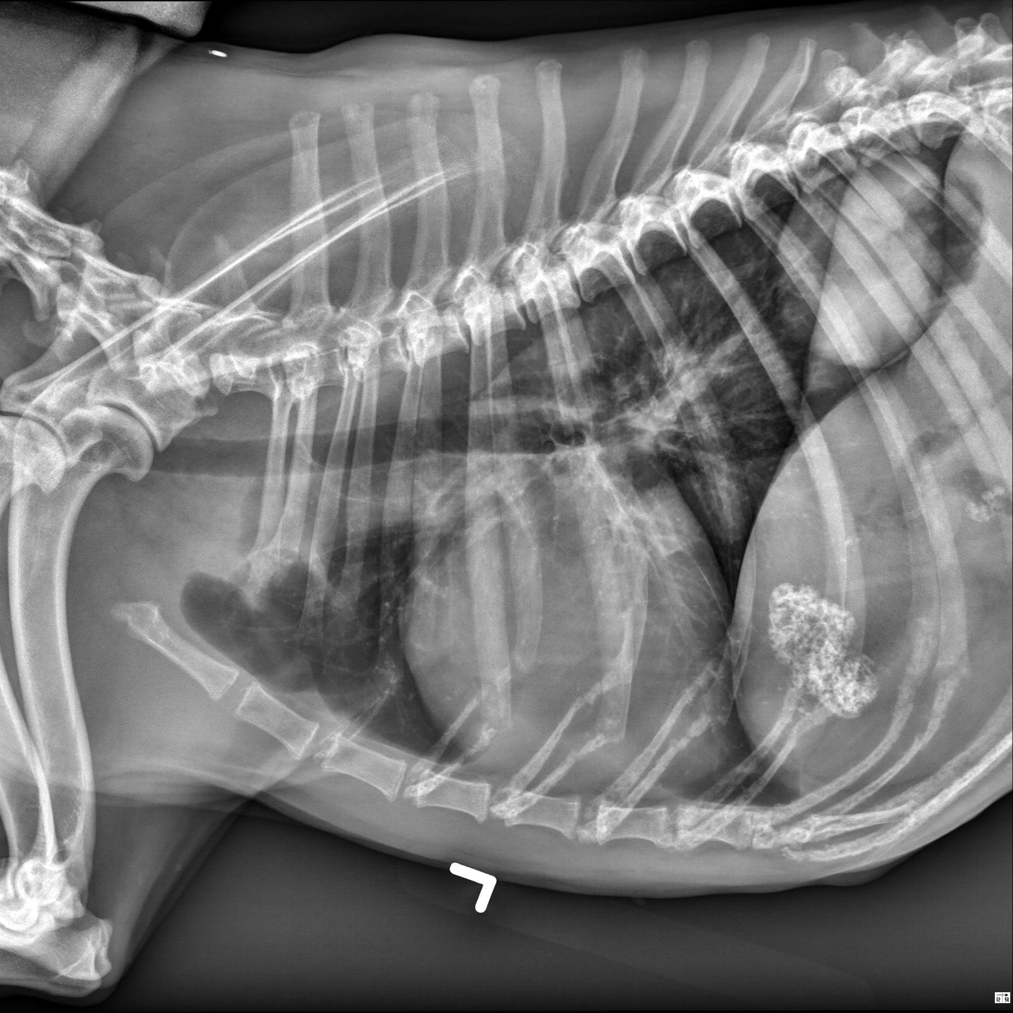
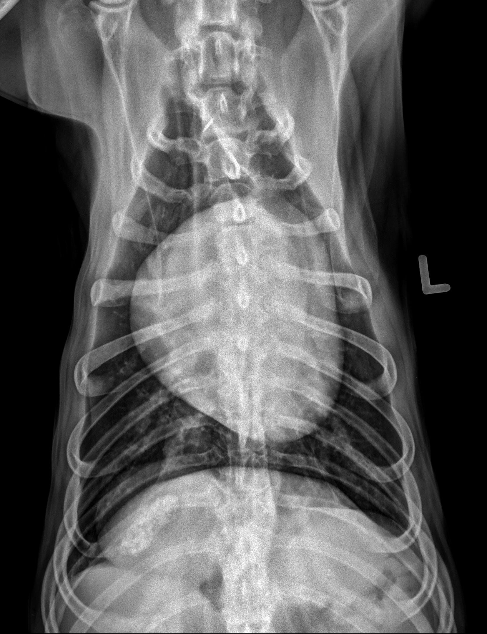
There is an oval, well-circumscribed, soft tissue opaque mass associated with the most caudal dorsal aspect of the right caudal lung lobe. This mass measures approximately 7.0 x 7.0 x 6.6 cm (length X height X width) based on the left lateral and VD views (without correction for magnification).
There are additional multifocal pinpoint mineral opaque foci throughout the lung fields, consistent with incidental pulmonary osteomata.
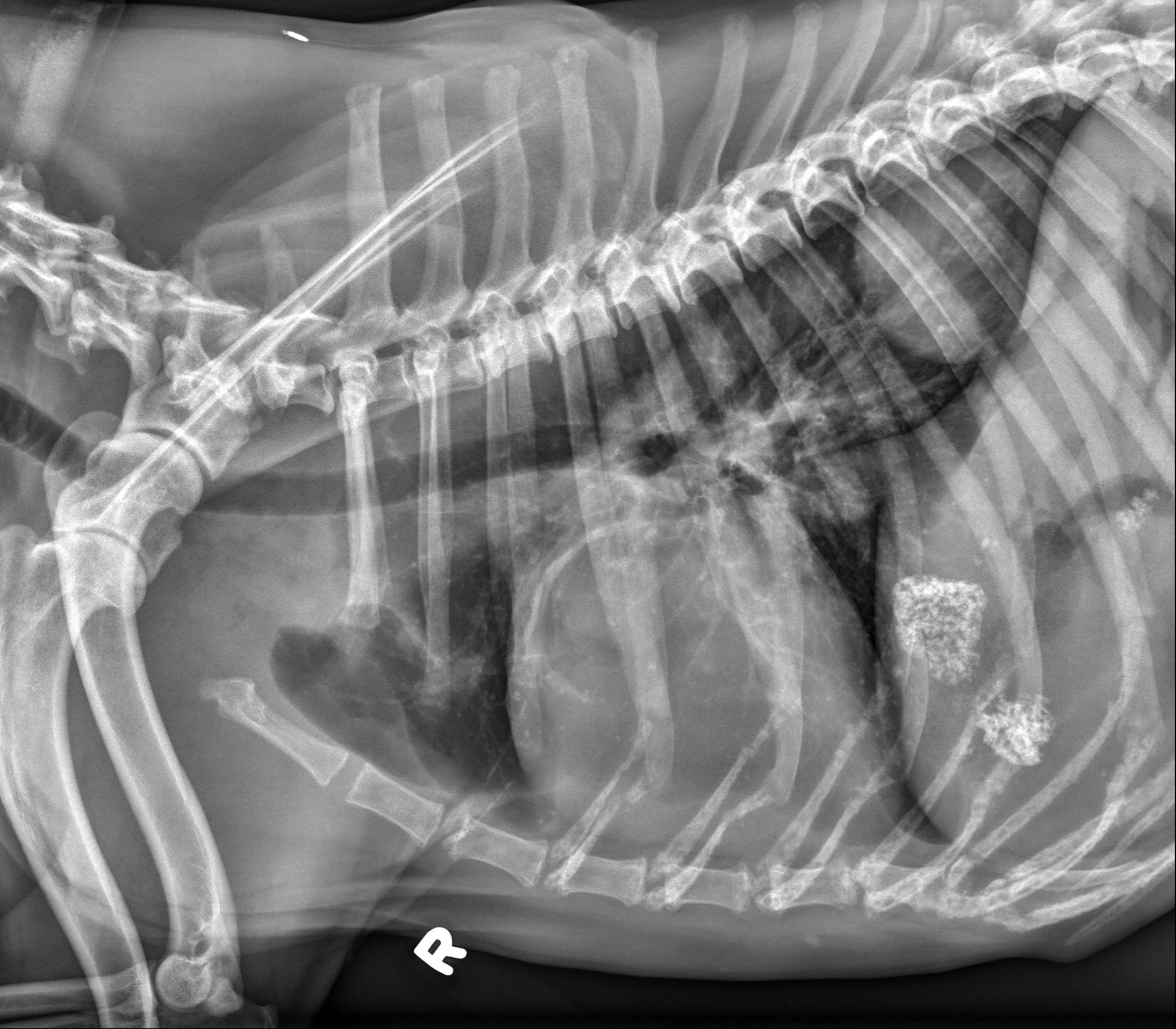
On the right lateral view, there is a curvilinear mineral opacity superimposed with the cranial dorsal aspect of the cardiac silhouette at the level of the third intercostal space which is not identified on the other views and likely represents either incidental mineralization of a pulmonary vessel or artifact.
An additional faint curvilinear soft tissue opacity is superimposed with the cranial dorsal thorax from the 1st-2nd pair of ribs on both lateral views, consistent with incidental plate like atelectasis.
The cardiovascular structures, mediastinal structures, pleural space, diaphragm, and thoracic wall are normal. There is incidental caudal thoracic spondylosis deformans.
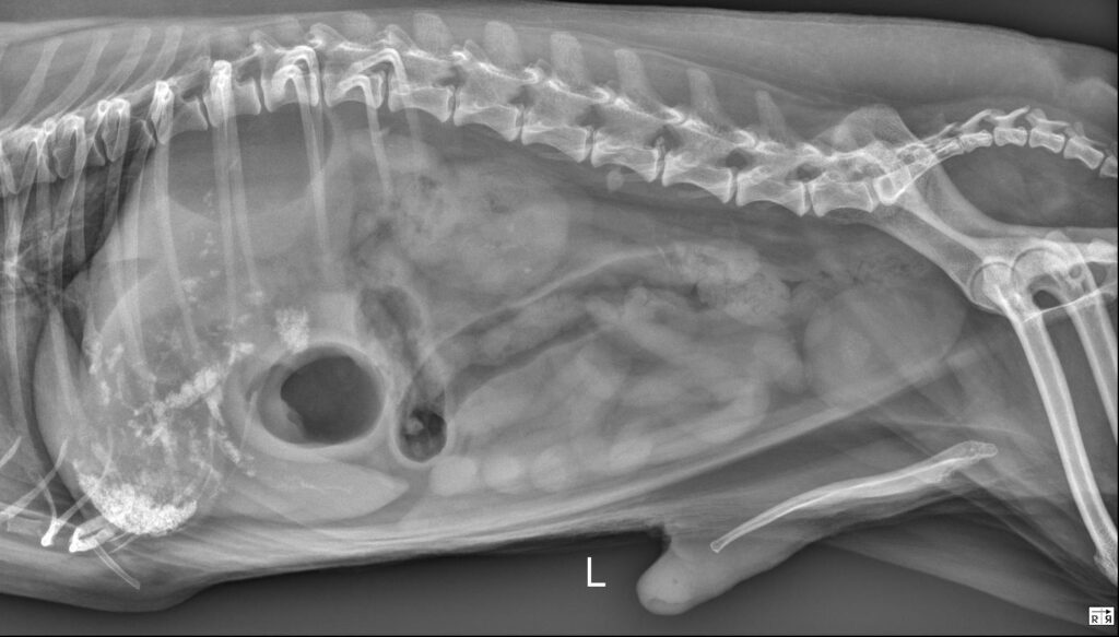
There are sharply marginated but irregularly shaped regions of heterogeneous mineral opacity superimposed with the right cranial aspect of the liver as well as the right cranial abdomen (superimposed with the expected location of the proximal descending duodenum on the lateral views and visible in the plane of the right 11th intercostal space on the VD view).
On both lateral views, there are faint branching gas opacities superimposed with the cranial abdomen which appear to be located within the hepatic parenchyma.
